
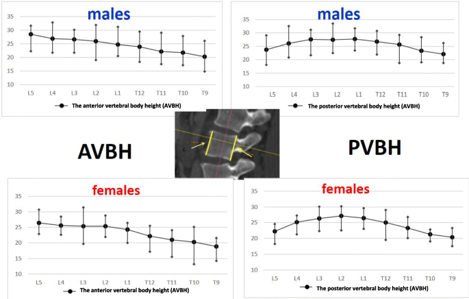
Vertebral Body Height Measurement
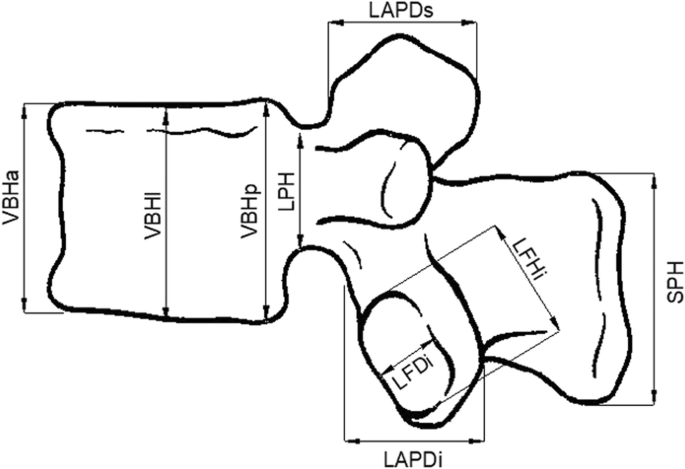
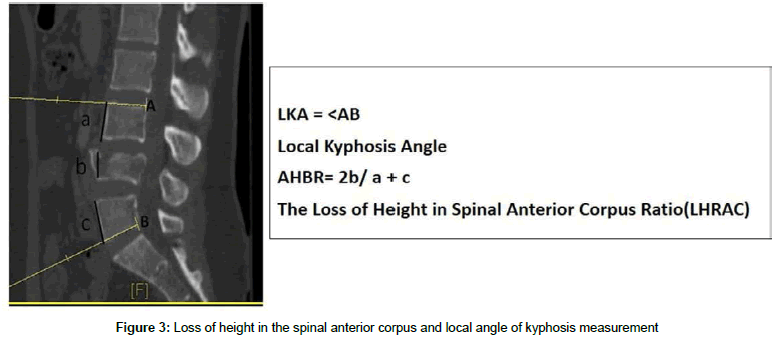
For compression fracture vertebral body height loss vbhl and kyphotic angle ka are two important imaging parameters for determining the prognosis and appropriate treatment. The objective of this study was to analyze the spinal dimensions of the ais spine in 3 d in vivo by measurement of the total length of the spine and height width depth ratios of the vertebral. The normal lower bound for relative change in measured vertebral heights ranged between 10 and 16 anteriorly and 11 and 19 posteriorly. Criteria for deformity should allow for this variation to avoid misdiagnosis. The average dorsal t12v body height was 2125164 mm in the control group and 2011149 mm in the lvf group. Anterior vertebral height remained the same from the third to the fifth vertebra but the posterior vertebral height decreased.
Previously researchers used different measurement methods to assess the degree of vertebral body collapse by using posterior wall height as the reference vertebral body compression ratio vbcr or the percentage of anterior height compression pahc which uses the mean height of segments adjacent to a healthy vertebral body as the reference value 8 11. The lvf group had significantly lower dorsal and ventral t12v body heights both p0001. Normal vertebral dimensions were found to vary from vertebra to vertebra. The average ventral t12v body heights were 1951154 mm and 1762195 mm respectively. Mean disc height in the lower lumbar segments was 116 18 mm for the l34 disc 113 21 mm for the l45 and 107 21 mm for the l5s1 level. This study used previous measurement methods to assess the degree of vbhl and ka compare and examine differences between various measurement methods and examine the correlation between relevant measurement parameters and intravertebral cleft ivc in the vertebral body.


Random Post
- komal jha body measurement
- gauhar khan body measurements
- aditi arya body measurement
- pori moni body measurement
- body fat measurement device omron
- rida isfahani body measurement
- jihyo twice body measurement
- best body measurement tracker
- ppt body measurement
- beautiful body measurement
- body measurement gzip
- fat and body measurements
- small size body measurements
- body measurement diagram
- flex lewis body measurements
- grace fit uk body measurements
- body measurement jquery
- zanilia zhao li ying body measurement
- stephanie rao body measurement
- body measurement chart
- kalki koechlin body measurement
- elephant body measurements
- jake gyllenhaal body measurements
- mike thurston body measurement
- diane lane body measurement
- stephen amell body measurements
- richa chadda body measurements
- laetitia casta body measurement
- rohit khandelwal body measurement
- body temperature measurement en español
- australian bra size measurement
- body measurement equivalents
- sofia hayat body measurements
- ideal body measurements cm
- objectives in taking body measurement
- victoria secret body measurement requirements
- body measurements for underweight
- eva mendes body measurement
- ruby may body measurements
- individual body measurements
- stefflon don body measurement
- analeigh tipton body measurement
- sarahs day body measurements
- the importance of body composition measurement at athletes and non athletes
- hareem shah body measurement
- body fat measurement auckland
- abc body measurements
- faf du plessis body measurement
- monalisa body measurement
- nancy jewel mcdonie body measurements
